High Voltage Qrs Complex
What Can Be The Reason For High Voltage Qrs Complex Quora
Qrs Interval Litfl Medical Blog Ecg Library Basics

Module 3 The Qrs Resus

Resting Ecg Showing Left Ventricular Hypertrophy High Qrs Voltage In Download Scientific Diagram
Ecg Fundamentals

High Voltage Cmc Ecg Masters

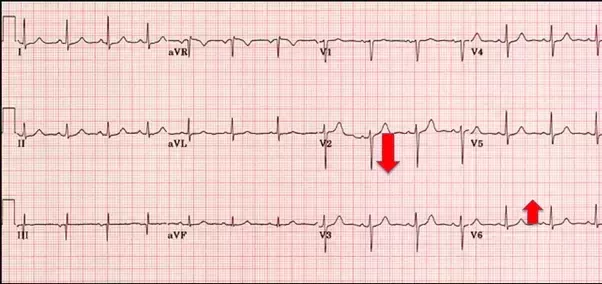
Refer to figure 6 panel a.
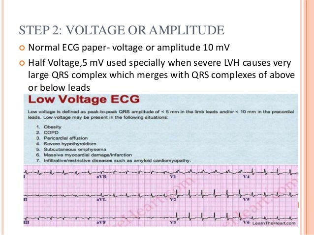
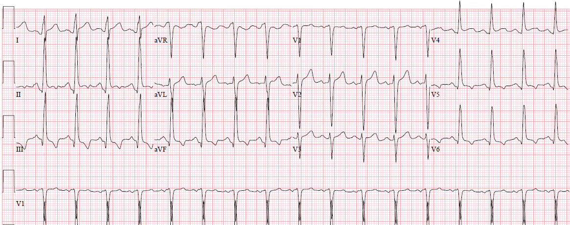
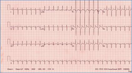
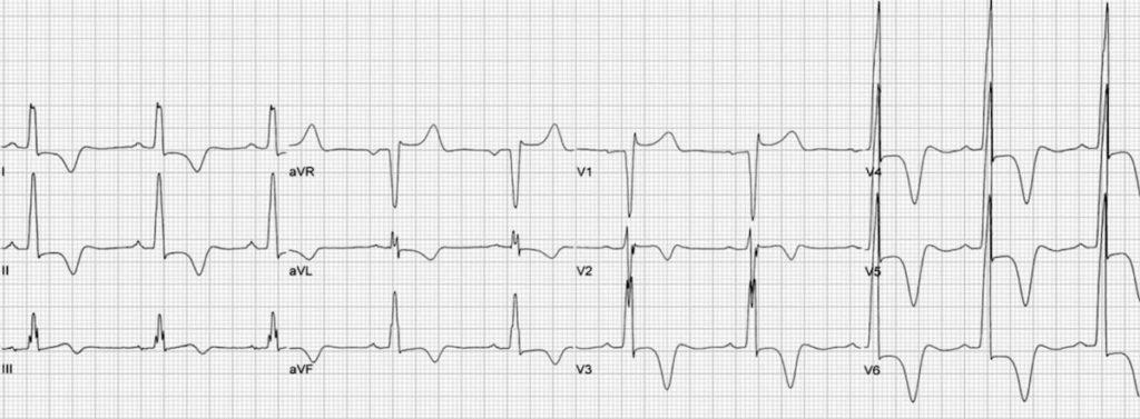
High voltage qrs complex.

Qrs Complex Thoracic Key

Physiological Lvh Dr S Venkatesan Md

Ecg Recorded Form Pda Dog Showed High Voltage Of Qrs Complexes Download Scientific Diagram

3rd Part Ecg Basics Qrs Complex Dr Salah Mabrouk Khallaf

Dr Smith S Ecg Blog Chest Pain Dynamic St Elevation And T Waves And High Voltage
Low Qrs Voltage Litfl Ecg Library Diagnosis
Basic Of Ecg And Easy Interpretation

Dr Smith S Ecg Blog History Of Hypertrophic Cardiomyopathy Hocm With Tachycardia And High Lactate

Conditions That Cause Abnormal Voltages Of The Qrs Complex

Ecg High Voltage Qrs St Depression And T Inversion Download Scientific Diagram
Dr Smith S Ecg Blog A Patient With Chest Pain And Dynamic St Elevation
Pediatric Ekg Interpretation

Dr Smith S Ecg Blog Page 34 Chan 6093258 Rssing Com
The Physiological Basis Of The Ekg Ppt Download

Dr Smith S Ecg Blog T Wave Inversions And Dynamic St Elevation

Guide To Understanding Ecg Artifact Acls Medical Training Medical Training Acls Medical Information

Electrocardiography Ecg And Normal Variants Elektrokardiyografide Normal Varyantlar Ekg Ecg Ankara Kardiyoloji Kalp Hastaliklari Mete Alpaslan Doktorekg Com

Ecg A Pictorial Primer
Https Encrypted Tbn0 Gstatic Com Images Q Tbn 3aand9gcrkwozoyzg2zcwzf2jsaxex9idsd2l 7vmzg0xm5ivpq19okooc Usqp Cau

Normal Ecg An Overview Sciencedirect Topics

What Is The Cause Of Low Voltage Unreadable Or Unclassified Alivecor Kardia Mobile Ecg Recordings The Skeptical Cardiologist
Hypertrophic Cardiomyopathy Hcm Litfl Ecg Library Diagnosis

Emergency Medicine News

Dr Smith S Ecg Blog Chest Pain And Convex St Elevation In Precordial Leads
Source : pinterest.com
