High Voltage Qrs Complex Causes
What Can Be The Reason For High Voltage Qrs Complex Quora
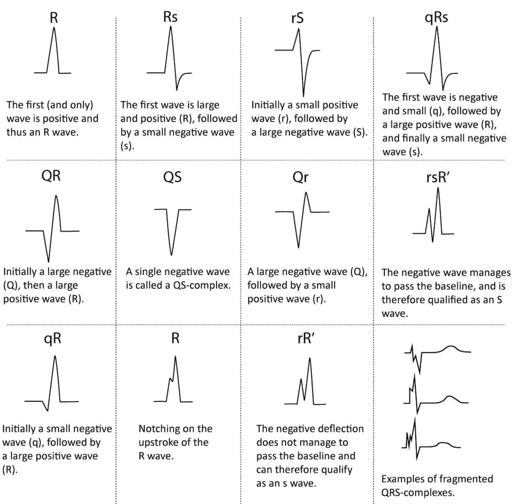
Qrs Interval Litfl Medical Blog Ecg Library Basics

Module 3 The Qrs Resus

Fragmented Wide Qrs Complex Fragmented Wide Qrs Complex In The Bundle Download Scientific Diagram

Ecg Interpretation Characteristics Of The Normal Ecg P Wave Qrs Complex St Segment T Wave Ecg Echo
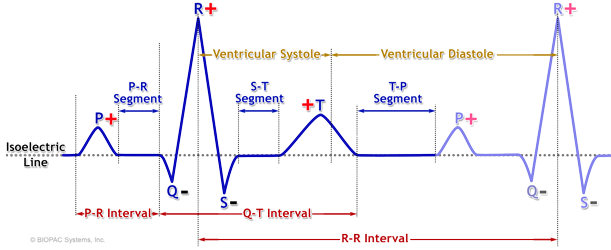
Qrs Amplitiude Respiratory Modulation Biopac

Again the spread takes place outside of the conduction system which is slow and causes widening of the qrs complex.
High voltage qrs complex causes.
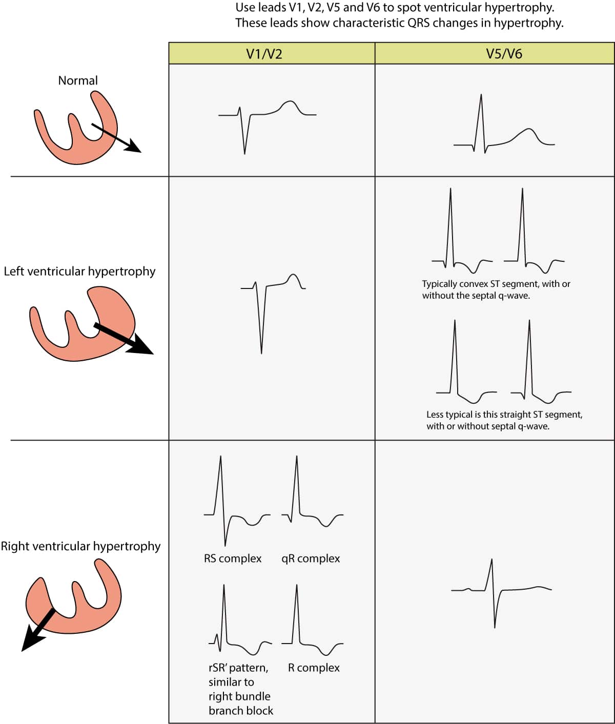
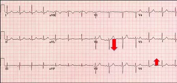
Ecg In Left Ventricular Hypertrophy Lvh Criteria And Implications Ecg Echo

Conditions That Cause Abnormal Voltages Of The Qrs Complex

Ecg A Pictorial Primer
Sinus Bradycardia Definitions Ecg Causes And Management Ecg Echo
Second Degree Heart Block Mobitz Type I Wenckebach P Waves And Qrs Complexes Are Normal But Pr Interval P Heart Blocks Pr Interval Ekg Interpretation

Pin By Jason Winter Ecg Educator On Cardiology Notes Nursing School Notes Nursing Notes Medical Student Study
Low Qrs Voltage Litfl Ecg Library Diagnosis

Rosh Review Nurse Cardiac Nursing Emergency Medicine

Pin By Aurora Zora On Medicine Cardiology Nursing Nurse Icu Nursing
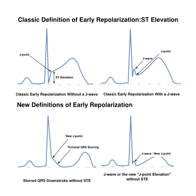
Early Repolarization Ecgpedia

Electrocardiography Hyperkalemia Ekg Ekg Rhythms

Ecg Primer For The Cath What Does A Tall R Wave In V1 Mean The Four Categories Approach Cath Lab Digest
Https Www Cardiacep Theclinics Com Article S1877 9182 19 30012 7 Pdf

Rosh Review Heart Blocks Cardiovascular System Ekg Rhythms

Pin By Jason Winter Ecg Educator On Ecg Ekg Study Memo Cards Cardiovascular System Ecg Rhythms Subarachnoid Hemorrhage

Med School Posts On Instagram Zoom In Ecg Findings Med School St Elevation Youtube Videos

7 Can T Miss Life Threatening Ecg Findings

Rosh Review Pediatric Patients Cardiac Nursing Nurse
Https Encrypted Tbn0 Gstatic Com Images Q Tbn 3aand9gcrkwozoyzg2zcwzf2jsaxex9idsd2l 7vmzg0xm5ivpq19okooc Usqp Cau
Hyperkalaemia Ecg Changes Litfl Ecg Library Diagnosis
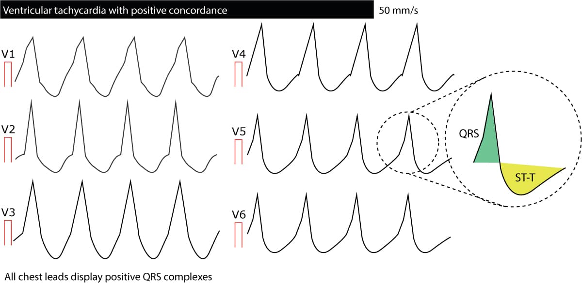
Ventricular Tachycardia Vt Ecg Criteria Causes Classification Treatment Management Ecg Echo

Pin By Anne Hollandsworth On Ecg Ekg Placement Ecg Rhythms Ekg

Pin On Atrial Flutter
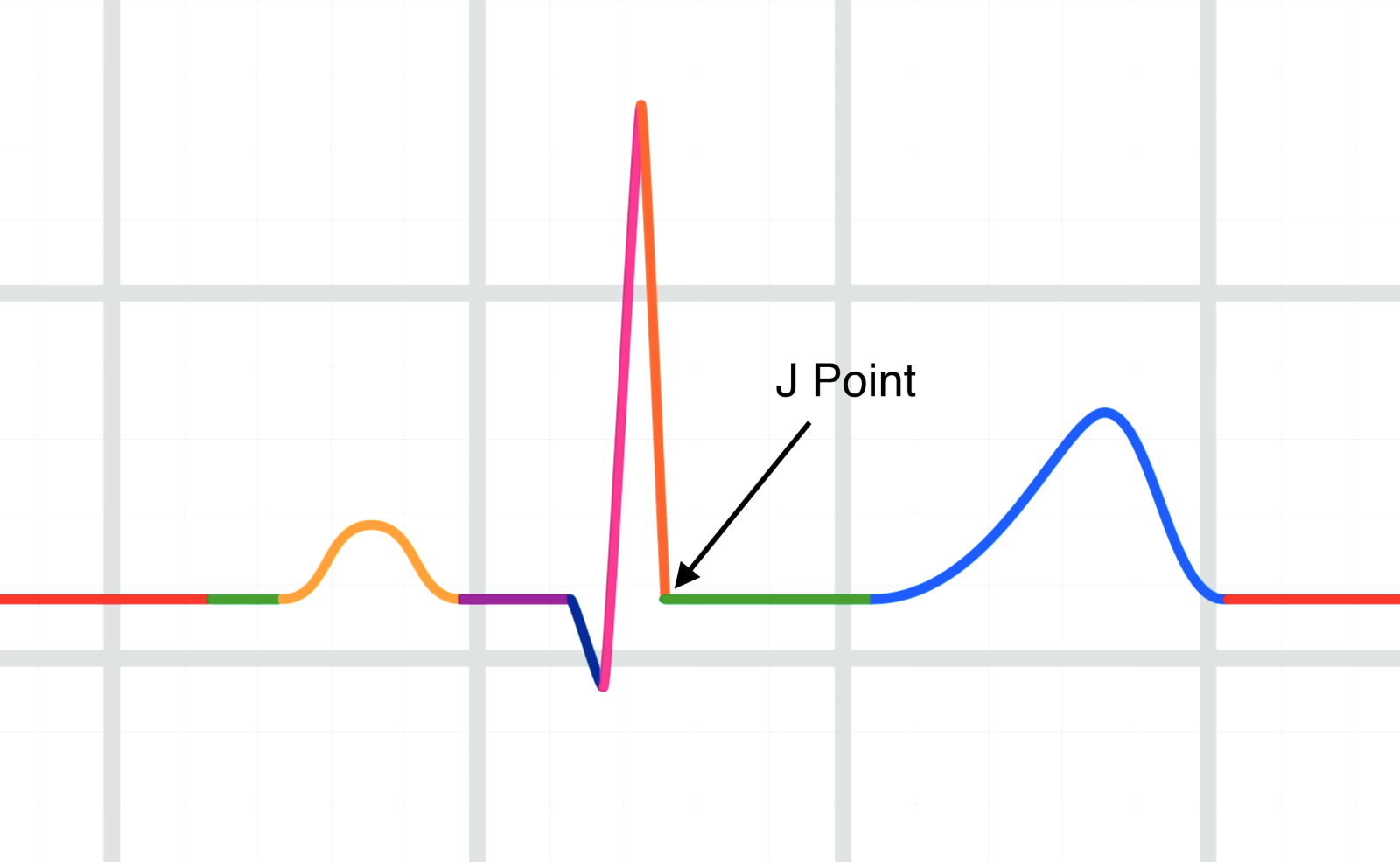
Lesson Title The J Point
Source : pinterest.com
